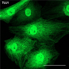
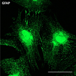
Differentiation ability of KHC: Nuronal cellKHCs can differentiate into neuronal cells.
Neuronal Cells Derived from KHCs
KHCs can regenerate damaged nervous systems within the body through this differentiation capability, offering a fundamental treatment for nervous system diseases.
bBHC's Research on Nervous System Diseases
Parkinson’s DiseaseParkinson's disease is a degenerative disorder of the central nervous system, primarily caused by damage to or loss of dopamine-producing neurons. It affects motor functions, leading to tremors, rigidity, bradykinesia (slow movement), and balance impairments. The causes of Parkinson’s disease are believed to involve a complex interplay of genetic and environmental factors, and current treatments focus mainly on symptom management.bBHC is using KHCs to advance the fight against Parkinson’s disease. When KHCs were intravenously administered to Parkinson’s model mice, motor neuron recovery was observed, and damaged brain tissue was also repaired.
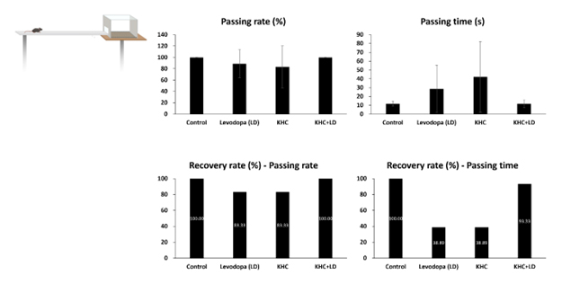
Balance Beam test - Passing rate, Passing time, Recovery rate
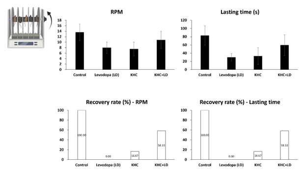
Rotaroad test - RPM, Lasting time, Recovery rate
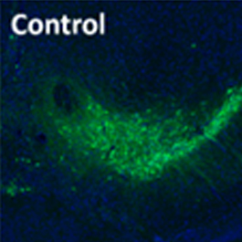
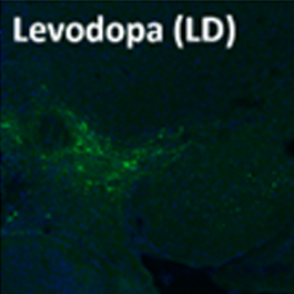
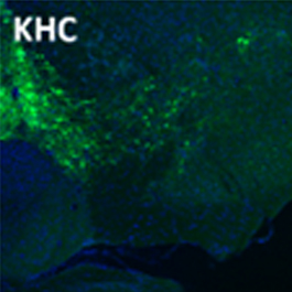
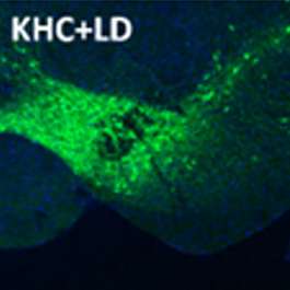
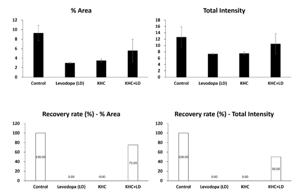
Dopaminergic neuron in substantia nigra of midbrain
The standalone administration of KHCs not only restores motor function to a level comparable to the most common Parkinson's disease treatment, levodopa but also regenerates damaged brain tissue—an ability levodopa lacks. Notably, the combined administration of KHCs and levodopa dramatically improves motor function and damaged brain tissue regeneration in Parkinson’s disease model mice.
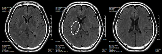
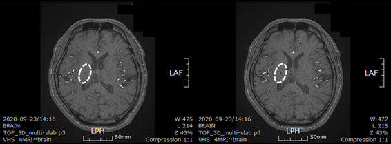
StrokeA stroke occurs when blood flow to the brain is blocked or reduced, damaging brain cells. Stroke is a medical emergency, with a golden window of 4.5 hours from symptom onset, and treatment within 24 hours is critical to prevent severe disabilities.A man who experienced a stroke (February 17, 2018) and underwent treatment without improvement, resulting in complete paralysis of the left side of his body, received minimally manipulated umbilical cord-derived mesenchymal stem cell transplantation approximately one month after the onset.
Brain CT images - 02/19/2018
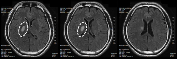
Brain CT images - 02/28/2018
Over one year following the transplantation, the patient gradually regained motor function, and by thirty months, most of the damaged brain tissue had regenerated.
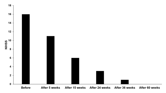
National Institute of Health Stroke Scale Score of Patient
Brain CT images - 30 months after first transplantation