COPYRIGHT@2020 BY bBHC Corporation Co.,Ltd.ALL RIGHT RESERVED
Stem Cell Treatment & Research Institution
Cell differentiation research:
To identify differentiation of nEPS for applying to any disease, we differentiated the nEPS into three germ layer cells of the ectoderm, the mesoderm, and the endoderm. Ectodermal cells formed neuronal cells, and mesodermal cells formed adipocyte, osteoblast and chondrocyte, and endoderm cells led to hepatocyte and pancreatic beta cells.
In the field of bioengineering, active research was conducted on applying stem cells in tissue engineering and differentiation mechanism of pluripotent stem cells like embryonic stem cell and induced pluripotent stem cell, and multipotent adult stem cells like bone marrow-derived stromal cell and adipose tissue-derived stem cell.
By the discovery of the differentiation potency of stem cells into diverse cells, there have been studies on signaling mechanisms and differentiation mechanisms that induce differentiation of stem cells into specific cells. There is also a tissue engineering attempt to create 3D tissues using stem cells. Stem Cell Treatment and Research Institute (STRI) has been studying the differentiation of umbilical cord-derived pluripotent stem cells into various body organ cells. Moreover, as well as studying new substances, proteins, culture environments and technologies in which such differentiation can occur more properly.
Ectodermal differentiation:
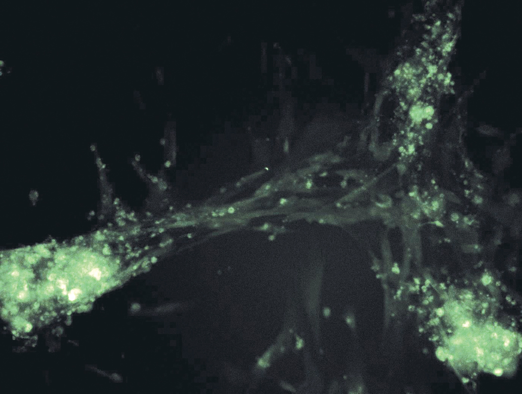
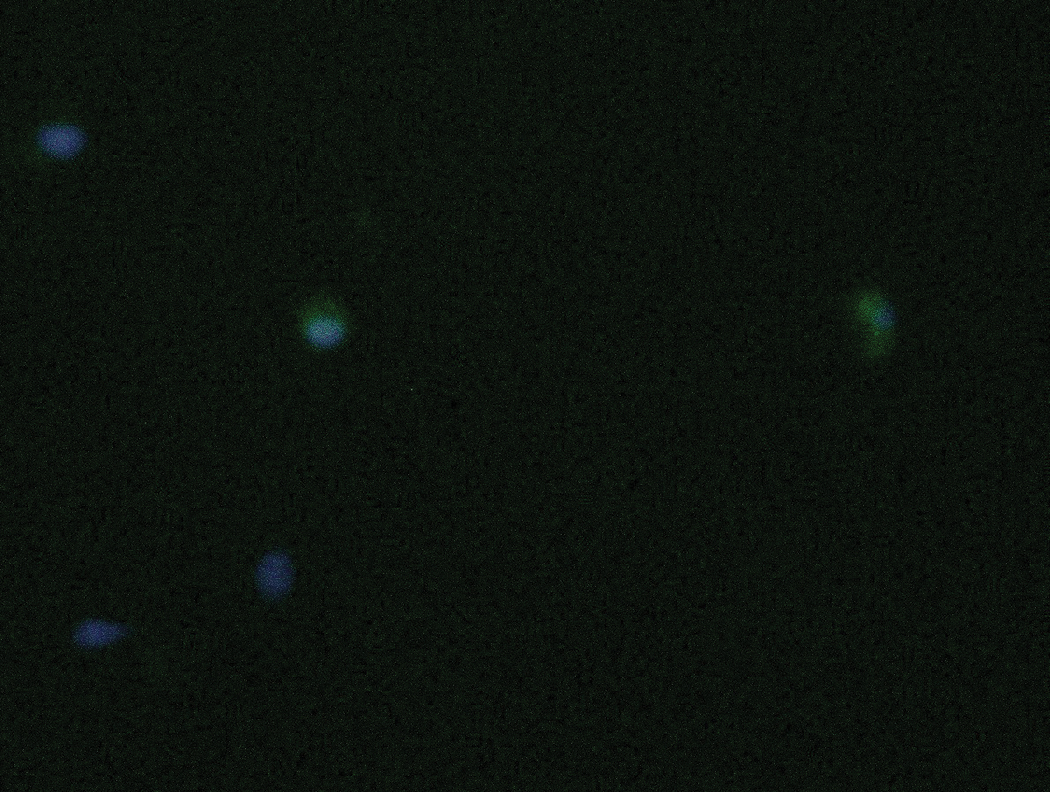
When the nEPS cell was differentiated into the ectodermal cell, it was differentiated into the shape of a nerve cell. Differentiation was verified by staining the differentiated cells with Nestin, which is only expressed in nerve cells. The differentiation was further verified as we were able to successfully merge Nesting and DNA-binding Hoechst dye together (Below – Neurocyte)
Endodermal differentiation:
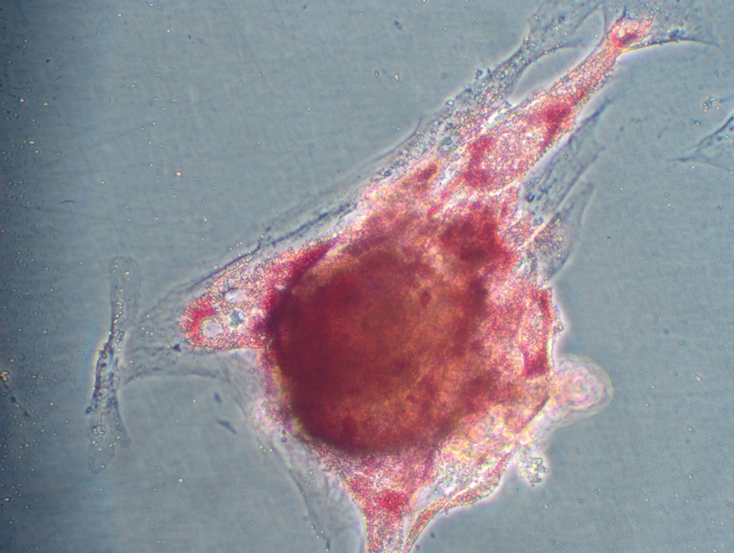
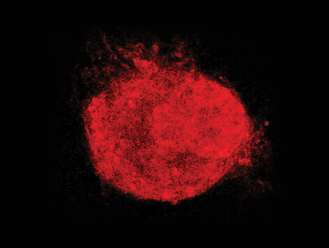
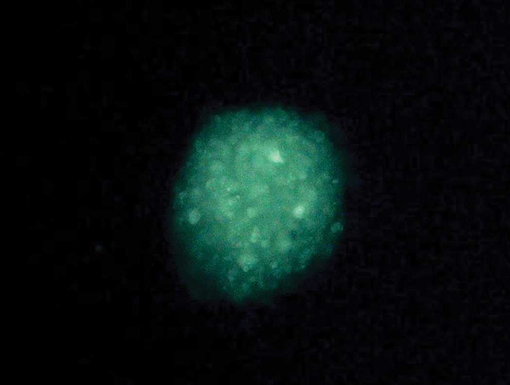
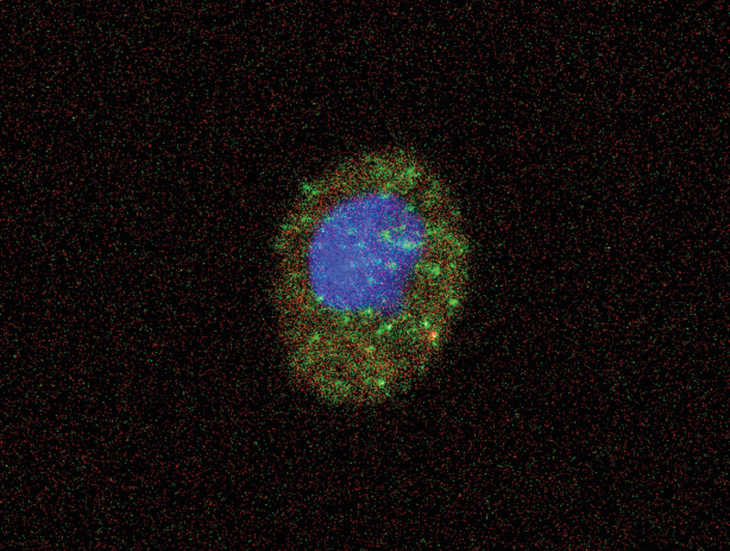
After differentiation of the nEPS cells into the liver cells, as the endodermal cells, a difference in the morphology of the cells was observed under microscope. Therefore, the differentiationwas verified by dyeing the cell with α-fetrotein, which is only expressed in liver cells. Merging the α-fetrotein expression result with DNA-binding Hoechst dye result successfully confirmed the differentiation of the nEPS cells into the liver cells. (Below – Hepatocyte)
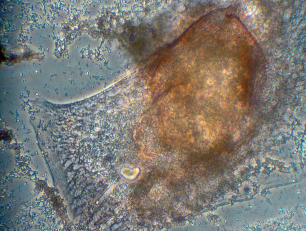
After nEPS cells were differentiated into pancreatic B-cell, there was a change in the shape of cellsdemonstrating the differentiation of nEPStopancreatic B-cell was changed. (Below - Pancreatic β-cell)
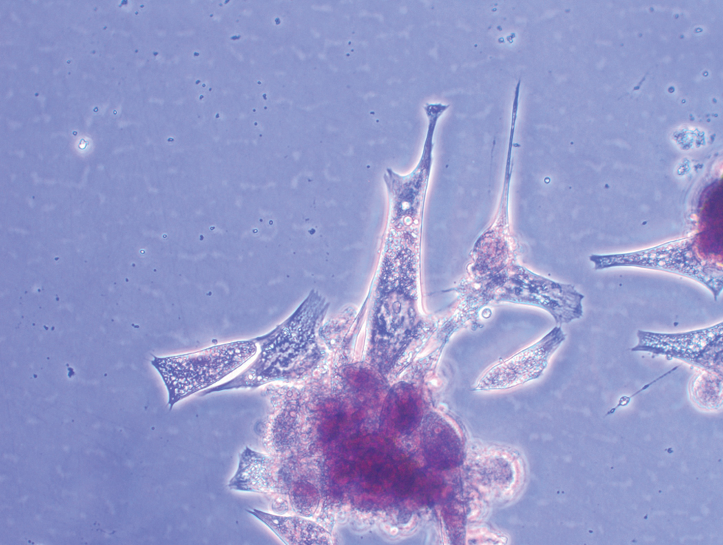
After nEPS cells were differentiated into kidney cell, there was a change in the shape of cellsdemonstrating the differentiation of nEPS to kidney cell. (Below – kidney cell)
Mesodermal differentiation:
ThenEPScellsasmesodermal cellsdifferentiated into cartilage and osteoblast cells. Cartilage cell consists of collagen and Polysaccharide polymer substance.
Differentiation into cartilage cell was verified by dyeing the cell with Alcian blue, which represents blue color through combination with polysaccharide having carboxyl sulfate unit at pH of 2.5.
After nEPS cells were differentiated into T cells,there was a change in the shape of cellsdemonstrating the differentiation of nEPSto T cell (Below – T cell).
After nEPS cells weredifferentiated into hematopoietic cells, there was a change in the shape of cellsdemonstrating the differentiation of nEPSto hematopoietic cell. (Below – hematopoietic cell)
After nEPS cells weredifferentiated intofat cells,there was a change in the shape of cellsdemonstrating the differentiation of nEPSto fat cell. (Below – fat cell)